
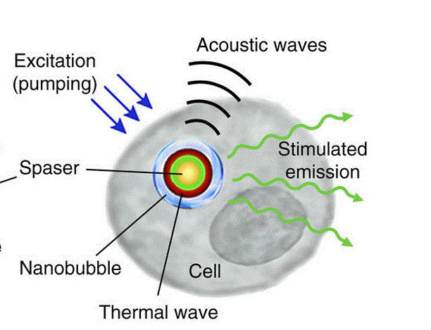

Spasers are energy-absorbing particles that release light. Spaser stands for “surface plasmon amplification by stimulated emission of radiation.” These tiny particles are a type of plasmonic nanoparticle. Lasers activate spasers, which can be targeted to cancer cells. Once internalized by cancer cells, spasers can selectively kill them.
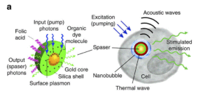
Galanzha and colleagues generated and tested spasers of different compositions to find ones that should be compatible for use in humans. The optimal spaser had a gold spherical nanoparticle at the core with a silica shell that incorporated the fluorescent dye uranine (Figure 1). These components have been used in medicine previously, suggesting this combination will also be safe for medical use. When exposed to an appropriately tuned laser, this spaser emitted heat that caused bubbles to form in the surrounding liquid. These bubbles amplified the intensity of the emission from the spaser while simultaneously reducing the width of the emission. This property of inducing the formation of bubbles should result in high-energy, deadly, and accurate spasers.
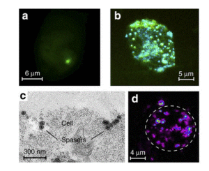
The next goal was to increase the selectivity of the spaser for cancer cells. A way that nanoparticles have been targeted to cancer cells is by incorporating folic acid. Cancer cells typically have higher numbers of receptors for folic acid than do normal cells. Thus, cells with high numbers of folate receptors selectively bound and internalized the spaser containing folic acid in the shell (Figure 2). Consistent with the potential toxicity of the spasers, laser irradiation of the cells that accumulated the spasers resulted in the formation of tiny bubbles inside the cells, which caused damage to the cell membrane and cellular fragmentation.
Spasers incorporated into cultured cells could be activated through a layer of human blood and spasers injected into the ears of mice could be activated within the tissue. Depending on the laser irradiation and the type of targeting molecules that are incorporated into the shell, these spasers could be useful as molecular detectors, diagnostic probes, or even targeted cell-destroying therapeutics.
Highlighted Article
Galanzha, E.I., Weingold, R., Nedosekin, D.A., Sarimollaoglu, M., Nolan, J., Harrington, W., Kuchyanov, A.S., Parkhomenko, R.G., Watanabe, F., Nima, Z., Biris, A.S., Plekhanov, A.I., Stockman, M.I., Zharov, V.P., Spaser as a biological probe. Nat. Commun. 8, 15528 (2017) doi: 10.1038/ncomms15528. PubMed
Related News Coverage
Spaser Can Detect, Kill Circulating Tumor Cells to Prevent Cancer Metastases. Georgia State University News (accessed 29 August 2017). News Story
Cite as: N. R. Gough, Spasers: Nanoparticles that Make Deadly Bubbles to Kill Cancer Cells. BioSerendipity (29 August 2017) https://www.bioserendipity.com/spasers-nanoparticles-that-make-deadly-bubbles-to-kill-cancer-cells/