

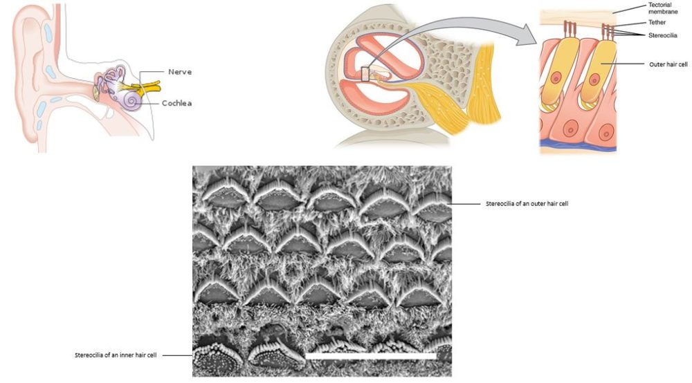
The top left panel shows a diagram of the parts of the inner ear. The top right shows a diagram of a section through the cochlea and an enlarged view of the parts of an outer hair cell. The stereocilia are the structures that resemble “hair.” The bottom image shows the three rows of outer hair cells and one row of inner hair cells in the cochlea of a mouse at birth. The white bar is a scale bar representing 15 micrometers.
Read about research into the inner ear:
N. R. Gough, A Key to Restoring Age-Related Hearing Loss. BioSerendipity (21 November 2018) https://www.bioserendipity.com/a-key-to-restoring-age-related-hearing-loss
Image Credits:
Top Left: Cancer Research UK [CC BY-SA 4.0 (https://creativecommons.org/licenses/by-sa/4.0)], via Wikimedia Commons
Top Right: OpenStax [CC BY 4.0 (https://creativecommons.org/licenses/by/4.0)], via Wikimedia Commons
Bottom: R. Hertzano, et al. PLoS Genet. 4: e1000207 (2008). https://doi.org/10.1371/journal.pgen.1000207